- m3.com 電子書籍
- 通常内視鏡観察による 早期胃癌の拾い上げと診断〔英語版〕

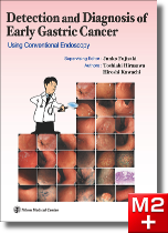
通常内視鏡観察による 早期胃癌の拾い上げと診断〔英語版〕
Junko Fujisaki (Supervising Editor) / Toshiaki Hirasawa , Hiroshi Kawachi (Authors) / 日本メディカルセンター
商品情報
内容
Having already gained some experience in endoscopy before I joined the Cancer Institute Hospital of JFCR, I was under the misapprehension that upper GI endoscopy was easy. At the Cancer Institute Hospital of JFCR, however, I saw lesions I had never seen before — such as gastritis-resembling cancer, minute gastric cancer, and 0-IIb lesions — being detected one after the other. I was astonished by the difference between my competence and that of the doctors at the Institute. When I asked one of them what the secret to finding gastric cancer was, he said that the lesion looked luminous. At first, I didn't understand what he meant. But once I had looked at enough lesions to get used to them, lesions started jumping out at me. And I understood it was what he meant when he said the lesion looked luminous. (from "Afterword")
> 通常内視鏡観察による 早期胃癌の拾い上げと診断〔日本語版〕
序文
This book originated when Dr. Toshiaki Hirasawa, one of the coauthors of this book, sent out quiz-style emails to residents and medical staff at Cancer Institute Hospital of JFCR. The email quiz had been developed as a teaching tool for use in resident training. A series of slides were attached that showed images of various unusual cases Dr. Hirasawa had worked with, as well as cases where lesions were missed.
In the first email distribution, the slides were not accompanied by the associated diagnoses. Instead the recipients were left to examine the images and come to a diagnosis on their own. The actual diagnoses were distributed a few days later. The clinical cases were all very instructive; they included typical cases, difficult-to-diagnose cases, cases where lesions were missed, and so on. The results were very positive. We staff doctors also found them beneficial.
In fact the emails proved so informative that Dr. Masahiro Igarashi, who was Director of the Department of Gastroenterology at the time, thought that it would be a good idea to turn the emails into a series of articles for a Japanese journal called Clinical Gastroenterology. The articles turned into a long-running serial, appearing in every issue and were subsequently collected and revised for inclusion in this volume.
The Cancer Institute Hospital of JFCR is a high-volume center, performing ESD for early cancer on almost 500 cases annually. All the cases are treated only after they have been examined at conferences. In fact, when surgeries are added in, it would be possible for a physician to study close to 1,000 cases of gastric cancer a year simply by attending the conferences. This book can be regarded as a comprehensive compilation of clinical cases that are experienced every day at the Cancer Institute Hospital of JFCR.
This book is so helpful, so packed with invaluable information, that readers will immediately find themselves looking at endoscopic images in a whole new way, starting with the very next endoscopy they perform. I was fortunate to have the opportunity to review all the clinical cases when they were written up for Clinical Gastroenterology. The data and the conclusions were so impressive and convincing that I was literally nodding non-stop while I looked over the material. In this book, you will find a wealth of practical information, including tips on detecting lesions,how to use endoscopes, and details on actual practice.
When we look at the clinical cases introduced in this book, we cannot avoid noticing that there are doctors today who are fully skilled in endoscopic submucosal detection (ESD), yet are unable to make an accurate diagnosis. On the other hand, there are doctors who have technical skill of ESD, yet totally incapable of finding and diagnosing them. This book attempts to solve these problems, making it a must-have for anyone who wants to master everything from detection to diagnosis using conventional endoscopy, which in turn is prerequisite that must be mastered before using magnifying endoscopy.
I have been watching as Dr. Hirasawa devoted all his energy to the creation of this book, so I cannot help feeling even more impressed by the result. As I have already mentioned, this extraordinary project had its genesis in our everyday work. Now it has turned into a single complete book. I hope that all doctors -- whether they are just starting out or have years of experience -- will find it helpful in their daily practice.
Finally, I would just like to congratulate Dr. Hirasawa on the completion of this book.
October 2016
Junko Fujisaki
Gastroenterological Medicine Department,
Cancer Institute Hospital of JFCR
And this book have just tranlated in English. I would like the doctors doing ESD all over the world to read this book.
March 2019
Junko Fujisaki
目次
Chapter Ⅰ Basic Knowledge in Gastric Cancer Detection
1 Normal gastric mucosa and gastritis
Normal gastric mucosa (Helicobacter pylori [H. pylori]-uninfected) / H. pylori-infected gastritis (H. pylori current infection and past infection)
2 Classification of gastric cancer and clinical characteristics
Classification of gastric cancer / Histogenesis and growth patterns of gastric cancer and clinical characteristics
Chapter Ⅱ How to Detect Gastric Cancer
1 Findings you need to consider in order to detect gastric cancer
Relationship with the background mucosa / Findings on the surface (cancerous area) / Findings on the margins
2 Factors and findings which make gastric cancer easy to miss
Sites that are easy to overlook / Lesions that are difficult to be found / Indigo carmine -- a magic potion that unveils hidden cancerss
Chapter Ⅲ Where's the Gastric Cancer? -- Detection and diagnosis
51 Cases are described.
Chapter Ⅳ Did You Find the Gastric Cancer? -- Answers and Diagnoses
便利機能
- 対応
- 一部対応
- 未対応
-
全文・
串刺検索 -
目次・
索引リンク - PCブラウザ閲覧
- メモ・付箋
-
PubMed
リンク - 動画再生
- 音声再生
- 今日の治療薬リンク
- イヤーノートリンク
-
南山堂医学
大辞典
リンク
- 対応
- 一部対応
- 未対応
対応機種
iOS 最新バージョンのOSをご利用ください
外部メモリ:111.9MB以上(インストール時:243.2MB以上)
ダウンロード時に必要なメモリ:447.6MB以上
AndroidOS 最新バージョンのOSをご利用ください
外部メモリ:72.9MB以上(インストール時:182.1MB以上)
ダウンロード時に必要なメモリ:291.6MB以上
- コンテンツのインストールにあたり、無線LANへの接続環境が必要です(3G回線によるインストールも可能ですが、データ量の多い通信のため、通信料が高額となりますので、無線LANを推奨しております)。
- コンテンツの使用にあたり、m3.com電子書籍アプリが必要です。 導入方法の詳細はこちら
- Appleロゴは、Apple Inc.の商標です。
- Androidロゴは Google LLC の商標です。
書籍情報
- ISBN:9784888753142
- ページ数:224頁
- 書籍発行日:2019年5月
- 電子版発売日:2019年6月12日
- 判:B5判
- 種別:eBook版 → 詳細はこちら
- 同時利用可能端末数:2
お客様の声
まだ投稿されていません
特記事項
※今日リンク、YNリンク、南山リンクについて、AndroidOSは今後一部製品から順次対応予定です。製品毎の対応/非対応は上の「便利機能」のアイコンをご確認下さいませ。
※ご入金確認後、メールにてご案内するダウンロード方法によりダウンロードしていただくとご使用いただけます。
※コンテンツの使用にあたり、m3.com 電子書籍(iOS/iPhoneOS/AndroidOS)が必要です。
※書籍の体裁そのままで表示しますため、ディスプレイサイズが7インチ以上の端末でのご使用を推奨します。


