- m3.com 電子書籍
- Kyoto Classification of Gastritis

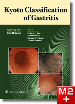
Kyoto Classification of Gastritis
Ken Haruma (Supervising Editor) / Mototsugu Kato , Kazuhiko Inoue , Kazunari Murakami , Tomoari Kamada (Editors) / 日本メディカルセンター
商品情報
序文
Preface
In this book, we will present and discuss the endoscopic findings required for diagnosis of gastritis in daily clinical practicewith a view to standardize the endoscopic diagnosis of gastritis in Japan.
Initially, the techniques used to diagnose gastritis were developed based on histopathological examinations of autopsied andresected stomachs. Diagnoses of gastritis were based on findings of erosion and inflammation of mucosa, as well ashypertrophy, atrophy, and intestinal metaplasia. Subsequently, the development of gastrocameras and fiberscopes made itpossible to observe the gastric mucosa with the naked eye, enabling physicians to diagnose gastritis by looking at opticallycaptured images of actual live tissue. This also made it possible to perform target biopsies; in other words, evaluation ofmicroscopic histopathological images with gastric biopsy was now added to surface observation with endoscopy. The netresult of these developments was a huge leap forward in the diagnosis and classification of gastritis.
The primary purpose for diagnosis of gastritis is evaluation of gastric mucosa, as this presents a potential risk of developinggastric cancer. Today, thanks to advances in endoscopic technology, it is now possible to observe gastric mucosa in minutedetail. Detection of even minor gastric mucosal changes is now commonplace, leading to a proliferation of endoscopicfindings and significantly increasing the complexity of gastritis classification.
However, with the discovery of Helicobacter pylori (hereinafter referred to as H. pylori) the major cause of gastritis becameclear. Taking this discovery into account, the Sydney System was created for classification and grading of gastritis. It waslater revised as the Updated Sydney System. Because it incorporates the impact of H. pylori infection, this breakthroughsystem effectively evaluates localization of gastritis and histopathological grading of gastritis, as well as category-specificendoscopic findings and diagnoses.
In Japan, the Kimura-Takemoto Classification of atrophic pattern and the Gastritis Study Group Classification remain thedominant standards in clinical practice. However, in order to bring the Japanese system in line with international practice,introduction of the Updated Sydney System was inevitable as it is the common available standard. Regardless, Japan alreadyhas a well-established and long-standing historical background in gastritis classification. It also happens to have a highoccurrence of gastric cancer. What Japan needs now is a gastric diagnosis system that evaluates the risk of gastric cancer.
From the 10th to 12th of May, 2013, I presided over the 85th Congress of the Japan Gastroenterological Endoscopy Society atthe Kyoto International Conference Center. Two main themes in gastritis diagnosis were discussed at this meeting. One washow to clarify the findings used to diagnose H. pylori-infected gastritis in standard endoscopic diagnostics in Japan whilestriving for greater objectivity and higher accuracy in endoscopic findings, as well as in histopathological findings based onthe Updated Sydney System. The other topic was to develop a method for grading gastritis that could evaluate a risk ofgastric cancer.
This book features images and accompanying descriptions that can be used as a basis for diagnosis of gastritis. Thephysicians who contributed to this book include many who participated in the discussions at the 85th Congress, as well asothers who are engaged in everyday practice of gastritis diagnosis. During the preparation of this book, we held dozens offace-to-face meetings and communicated constantly via the Internet to ensure that we were all agreed on the classificationsdescribed herein.
As both supervising editor and contributing author, I look forward to seeing this classification system validated in clinicalcases, and hope that it will be upgraded regularly, and evaluated internationally.
May 2017
Ken Haruma
Professor, General Internal Medicine 2,
General Medical Center,
Kawasaki Medical School
目次
Chapter 1 History of the Classification of Gastritis
Chapter 2 Endoscopic Findings of Gastritis
1. Introduction
(1) H. pylori-uninfected gastric mucosa = Normal stomach
(2) Currently H. pylori-infected gastric mucosa = Chronic active gastritis (CAG)
(3) Previously H. pylori-infected gastric mucosa (natural disappearance of H. pylori after eradication or advanced atrophy)= Chronic inactive gastritis (CIG)
(4) Changes in the gastric mucosa caused by drugs
2. Specific Discussions
Atrophy / Intestinal metaplasia / Diffuse redness / Spotty redness / Mucosal swelling / Enlarged folds and tortuous folds / Nodularity / Foveolar-hyperplastic polyp / Xanthoma / Depressive erosion / Regular arrangement of collecting venules (RAC) / Fundic gland polyp / Red streak / Raised erosion / Hematin / Corpus erosion / Patchy redness / Map-like redness / Multiple white and flat elevated lesions / Cobblestone Mucos
Chapter 3 Endoscopic Findings for Risk Stratification of Gastric Cancer
1. Description
(1) Relationship between gastric cancer and background gastritis
(2) Endoscopic findings related to the risk of gastric cancer
(3) Scores for endoscopic findings related to the risk of gastric cance
2. Clinical Cases
Chapter 4 Recording Endoscopic Findings of Gastritis
1. Description and Clinical Cases
(1) Basic method for entering data
(2) Entering endoscopic findings of gastritis according to clinical cases
2. Check Sheet for Background Gastric Mucosa in Endoscopy
3. Endoscopic Diagnosis and Classification of Chronic Gastritis That Conforms to Histological Diagnosis
(1) Diagnosis policy of chronic gastritis
(2) Diagnosis of presence/absence and activity of chronic gastritis
(3) Diagnosis of atrophy
(4) Consistency with pathological diagnosis
便利機能
- 対応
- 一部対応
- 未対応
-
全文・
串刺検索 -
目次・
索引リンク - PCブラウザ閲覧
- メモ・付箋
-
PubMed
リンク - 動画再生
- 音声再生
- 今日の治療薬リンク
- イヤーノートリンク
-
南山堂医学
大辞典
リンク
- 対応
- 一部対応
- 未対応
対応機種
iOS 最新バージョンのOSをご利用ください
外部メモリ:63.0MB以上(インストール時:137.1MB以上)
ダウンロード時に必要なメモリ:252.0MB以上
AndroidOS 最新バージョンのOSをご利用ください
外部メモリ:30.5MB以上(インストール時:76.2MB以上)
ダウンロード時に必要なメモリ:122.0MB以上
- コンテンツのインストールにあたり、無線LANへの接続環境が必要です(3G回線によるインストールも可能ですが、データ量の多い通信のため、通信料が高額となりますので、無線LANを推奨しております)。
- コンテンツの使用にあたり、m3.com電子書籍アプリが必要です。 導入方法の詳細はこちら
- Appleロゴは、Apple Inc.の商標です。
- Androidロゴは Google LLC の商標です。
書籍情報
- ISBN:9784888759267
- ページ数:236頁
- 書籍発行日:2017年9月
- 電子版発売日:2018年8月24日
- 判:B5判
- 種別:eBook版 → 詳細はこちら
- 同時利用可能端末数:2
お客様の声
まだ投稿されていません
特記事項
※今日リンク、YNリンク、南山リンクについて、AndroidOSは今後一部製品から順次対応予定です。製品毎の対応/非対応は上の「便利機能」のアイコンをご確認下さいませ。
※ご入金確認後、メールにてご案内するダウンロード方法によりダウンロードしていただくとご使用いただけます。
※コンテンツの使用にあたり、m3.com 電子書籍(iOS/iPhoneOS/AndroidOS)が必要です。
※書籍の体裁そのままで表示しますため、ディスプレイサイズが7インチ以上の端末でのご使用を推奨します。


